Light Microscopy (LM) is a very broad range of methods that use optical lenses to create magnified images of small objects. Three main types of light microscopes can be discerned: Stereo microscope, compound microscope, and confocal laser scanning microscope.
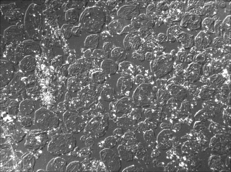
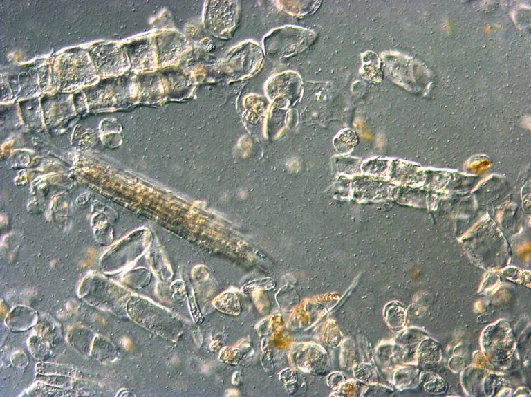
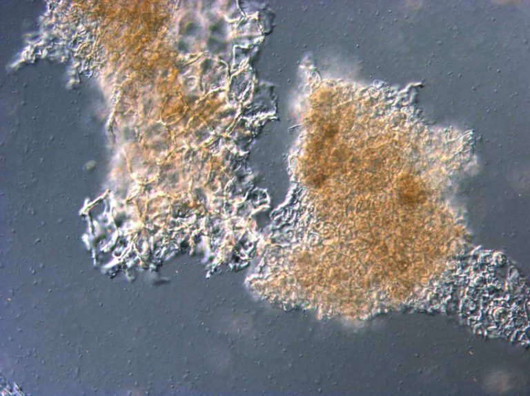
Differential Interference Contrast Light Microscopy image of a commercial tomato ketchup. Here this method highlights cell walls and swollen starch grains. The same area has been imaged with LM-BF and LM-CPOL as well.
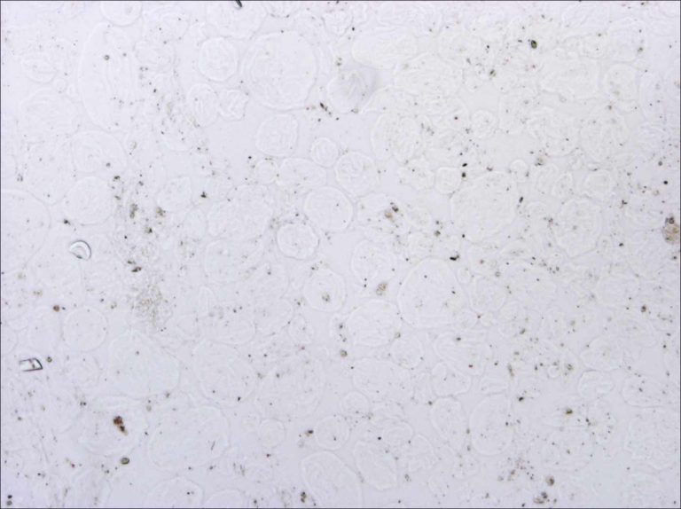
Bright Field Light Microscopy image of a commercial tomato ketchup. Next images show the same area viewed with different methods (LM-DIC and LM-CPOL). LM-BF is the basis imaging mode of light microscopy. Without the use of dyes, LM-BF images often have poor contrast.
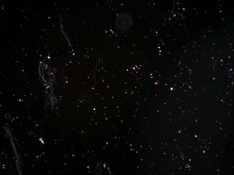
Circular Polarised Light Microscopy image of a commercial tomato ketchup (same area also imaged with LM-BF and LM-DIC). In this sample the lycopene crystals and cell wall material are highlighted by this method.
Stereo microscope
This instrument is also called dissection microscope and macroscope. The stereo microscope delivers high magnifications of 3D surfaces. They are typically used by entomologists, by watch makers, and for quality checks in fine productions. In research laboratories, stereo microscopes are often used for sample preparation for other methodologies, such as scanning electron microscopy.
Stereo microscopes can also be used to produce high resolution images of features that are just too small to image with a regular camera, such as details of insects, plants, (non-)woven tissues, and powders. The word stereo is a bit misleading, as it refers to the binocular viewing providing a 3D view for the observer, but the digital images are generally made without such 3D effect. Stereo microscopy is often complementary to Scanning Electron Microscopy, adding colour data, but much less focal depth.
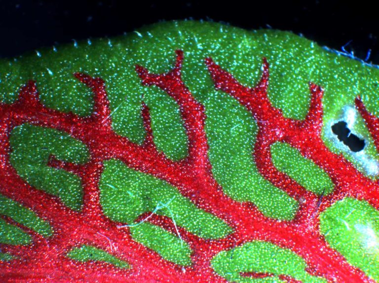
Stereo Microscope image of a plant leaf with herbivory damage
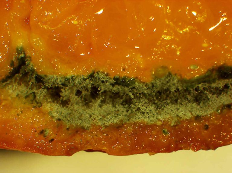
Stereo Microscope image of Mandarin Orange with fungal infection.
Stereo Microscopy photograph of an image detail shown on a smart phone screen. As can be seen here, this phone screen consists of red, blue, and green LED lights. Each neighbouring 1 red, 1 blue, and 2 green dots represent one pixel. Each pixel can create any color in any shade when seen from a distance.
The best magnifying glass is a stereo microscope with a good camera
Compound microscope
This is the light microscope sensu stricto. In many cases a thin section or a droplet of the sample is situated between two glass slides, the object slide and the cover slip, allowing light to travel from the light source through the sample into the lenses. The most straightforward light microscopy is called Bright Field microscopy, but besides that there are many different setups possible, all providing specific advantages. Here a number of types of light microscopy are listed:
Differential Interference Contrast Microscopy (LM-DIC or Nomarski) image of Chrysanthemum cut flower pith tissue revealing cell wall pits at high contrast.
Differential Interference Contrast Microscopy (LM-DIC or Nomarski) image of Cabbage hotbreak puree.
Differential Interference Contrast Microscopy (LM-DIC or Nomarski) image of Cabbage coldbreak puree.
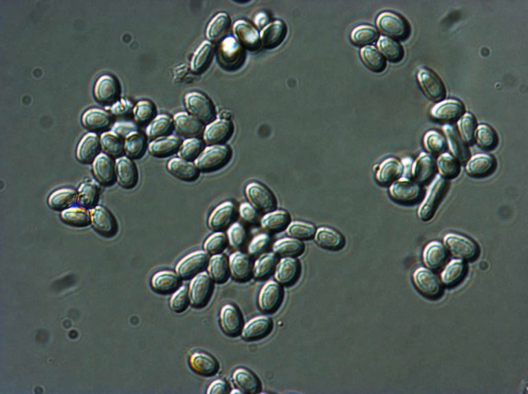
Differential Interference Contrast Microscopy (LM-DIC or Nomarski) image of fungal spores in citrus fruit. Image Width is 84µm.
Bright Field microscopy (LM-BF): The image shows the features on a light background, visualising microstructures as they are. Dyes can be used to identify specific features or to provide suitable contrast.
Dark Field microscopy (LM-DF): The image shows illuminated features on a dark background. This method is used to highlight features in a rather empty medium, such as fibrils, fine particles etc, that would have much less contrast with bright field microscopy.
Phase contrast microscopy (LM_PC). Light transmitted through a sample can be of different phases, depending on the materials and interfaces it passed. Phase contrast microscopy applies an optical trick to highlight phase changes, thereby providing contrast to samples. This method can be used to better view emulsions, dispersions, and cellular components.
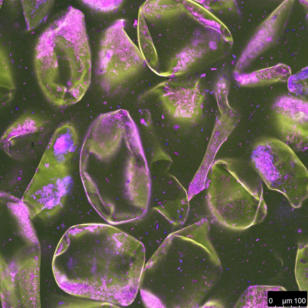
Fluorescence microscopy (LM-FL): Some materials emit light of a different wavelength than the illuminating source. This phenomenon is called fluorescence. In this case materials or tracing dyes and tracing particles can be localised, by irradiating the sample with a wavelength that is blocked for the eye, thus creating an image from the fluorescence light only. This method has many applications, mainly for localisation of (bio) molecules and/or to obtain better contrast at high resolution imaging.
Polarised light microscopy (LM-POL), incl circular polarised light microscopy. With this method the light from the light source is polarised and light that is transmitted through the sample is polarised into the opposite direction, leaving a black field in absence of sample material. If the sample contains ordered structures, such as crystals or aligned fibrils or lamellae, the polarisation of the light is changed. Only the light with changed polarisation is transmitted to the image, resulting in illuminated features on a black background.
Differential Interference Contrast microscopy (LM-DIC), also called Nomarski microscopy. This method could be practically regarded as a refined version of phase contrast microscopy. Phase contrast microscopy can yield blurred images due to many superimposed highlighted interfaces, especially in relatively thick samples. LM-DIC only highlights interfaces in rather thin optical sections of the sample, thereby yielding much clearer images than LM-PC, with an artificial light-and-shadow 3D-effect. LM-DIC requires a considerable adaptation to a standard microscope, but if this method is installed, it is often used because of the high contrast in untreated samples without use of dyes.
Consistence has all LM methods mentioned above. Often, we combine several imaging modes to address your research questions. Very often we also include detailed scanning electron microscopy and CSLM to obtain high resolution data of a limited number of ‘cornerstone’ samples for deep insights, followed by the much faster LM-imaging of higher numbers of intermediate samples to obtain the full overview. Such a multidisciplinary imaging approach reduces the risk of overlooking crucial features and helps to create efficient and excellent interpretation.
A special service of Consistence is to translate multidisciplinary findings into schematic drawings, which in turn can be utilised to communicate a new hypothesis, patent, or processing insight. Light microscopes can be booked at Consistence for use by third parties. Consistence also provides help in optimising light microscopy setups in other R&D laboratories.
Contact us
To visit our laboratory, send a sample or learn what we can do for you.
email: info at consistence.nl
tel: +31 6 4468 1439
address: Fennaweg 53, 2991 ZA Barendrecht, The Netherlands.
Our aim is to provide world class microstructural insights and we are happy to receive feedback. Contact us for more information if this page is relevant to your research. Free use of the images and content of this webpage is permitted with reference to Consistence. Contact us for full resolution images.