Scanning Electron Microscopy

SEM

Scanning Electron Microscopy (SEM) is a method where a focussed beam of electrons is scanned over the sample surface. The recorded signal is scanned onto a screen or into a digital image. A conventional SEM requires a high vacuum sample chamber, where the electron bundle interacts with the sample. Low Vacuum and environmental SEM instruments are available as well.

Detection signals

Several types of electron sources have been developed to deliver optimal imaging results, either in costs or in image resolution. Different types of signals are generated by the electron bundle, of which the most used are mentioned here:

Backscattered Electrons

If the electron beam hits the sample, some of these primary electrons are scattered back into the SEM chamber.

Secondary Electrons

The electron beam hits the sample and causes other electrons to emit from the sample.

X-rays

This is a type of fluorescence, where electrons with higher energy generate X-rays with element-specific lower energies, resulting in spectral information that can be applied to localise and quantify the different components of materials based on atom composition.

Cryo-SEM is a key method for Soft Matter research

Cryo-SEM

Scanning electron microscopy is very useful for high resolution characterisation of sample surfaces, both in life sciences and in material sciences and related technologies. These surfaces could be natural surfaces or new surfaces made for analysis, such as cross sections and fracture faces.

Cryo Scanning Electron Microscopy (cryo-SEM), a special method using a deep cooled stage, allows imaging of hydrated specimens by freezing the samples to very low temperatures (below -100°C) where solid water is stable even in vacuum.

Cryo-SEM is very useful to analyse wet samples, in either three ways:

Stereo cryo-SEM image of two plant mites. Use red-green glasses to obtain 3D-view. Image by Frank Nijsse, www.Consistence.nl

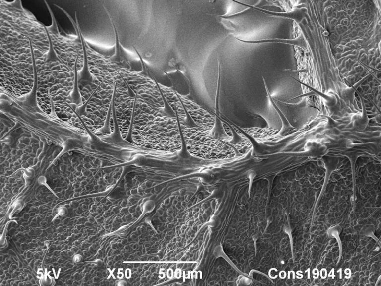
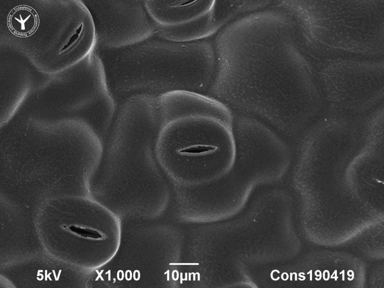
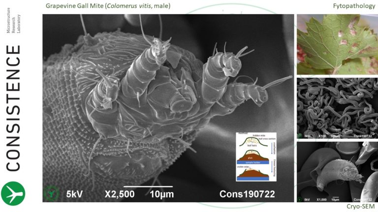
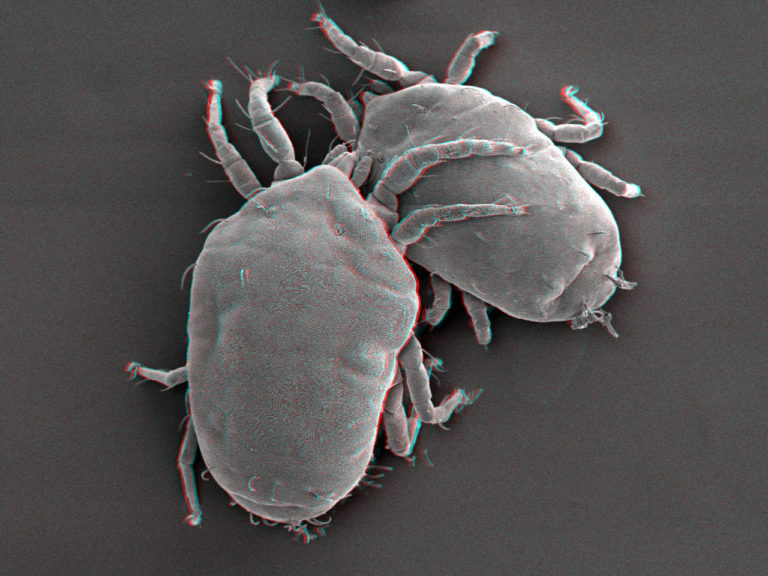
1. Cryo-SEM imaging of natural surface, such as high resolution imaging of insect or leaf surfaces.

Cryo-SEM fracture image of cabbage (Brassica Oleracea) leaf cross section. Image width is 1200 µm. Photo by Jaap Nijsse, Consistence Microstructure Research Laboratory.

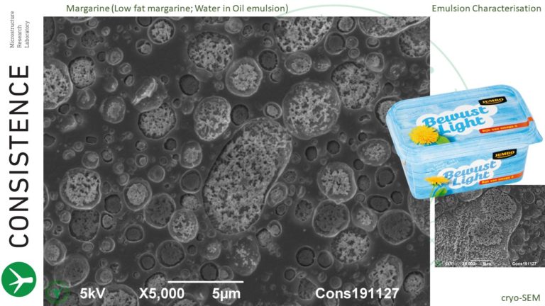
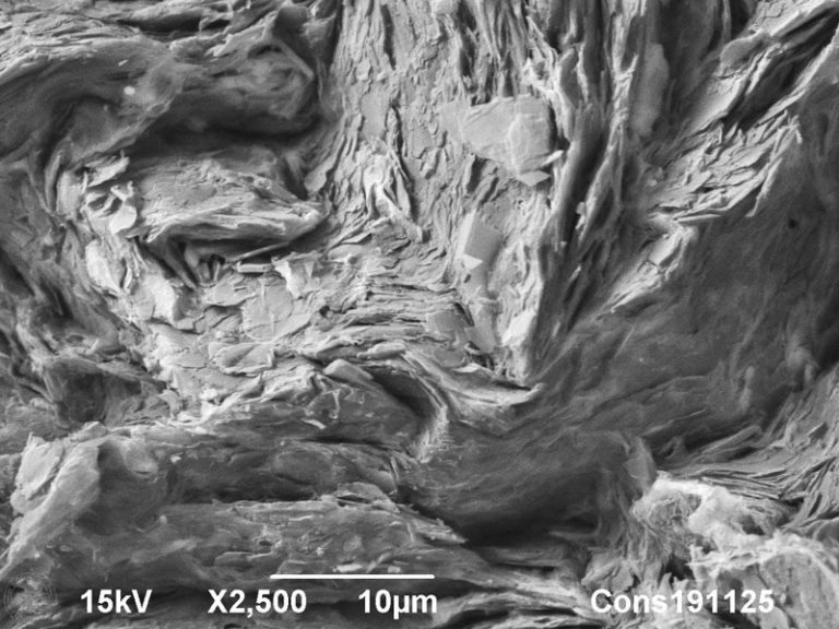
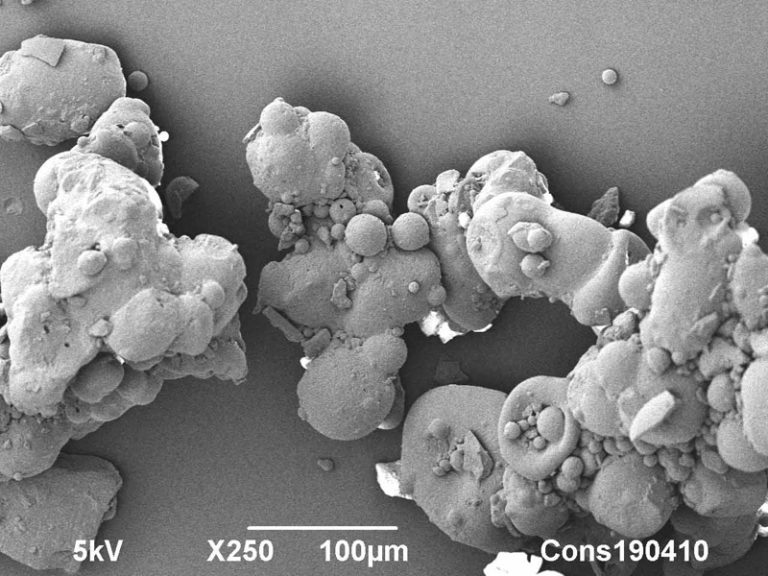
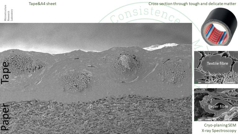
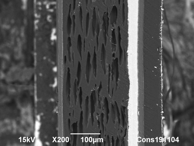
2. Cryo-SEM imaging of fracture induced surfaces through samples, revealing internal structures (Freeze-fracturing SEM). Often a fracture proceeds over interfaces and therefore this method is very useful to study interfaces in complex materials, such as foams, oil/water interfaces in emulsions, and membranes in living cells.

Cryoplaning SEM image of cross section through fibrous particle in cabbage puree. Image width is 120 µm. Photo by Jaap Nijsse, Consistence Microstructure Research Laboratory.

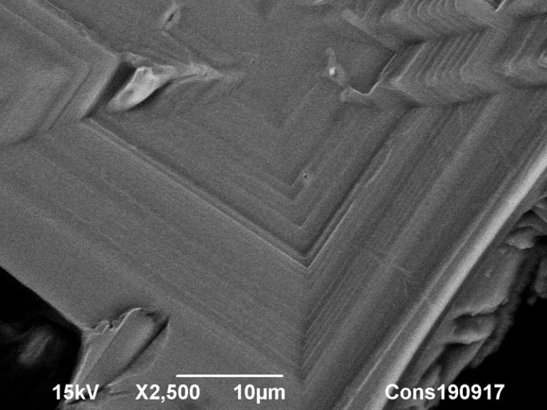
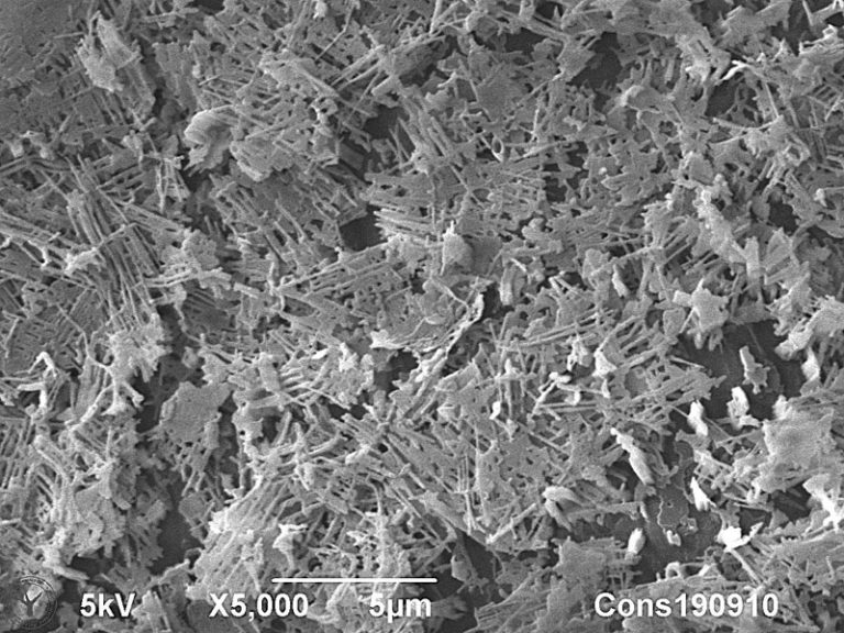
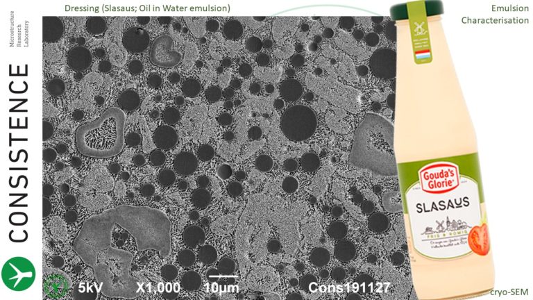
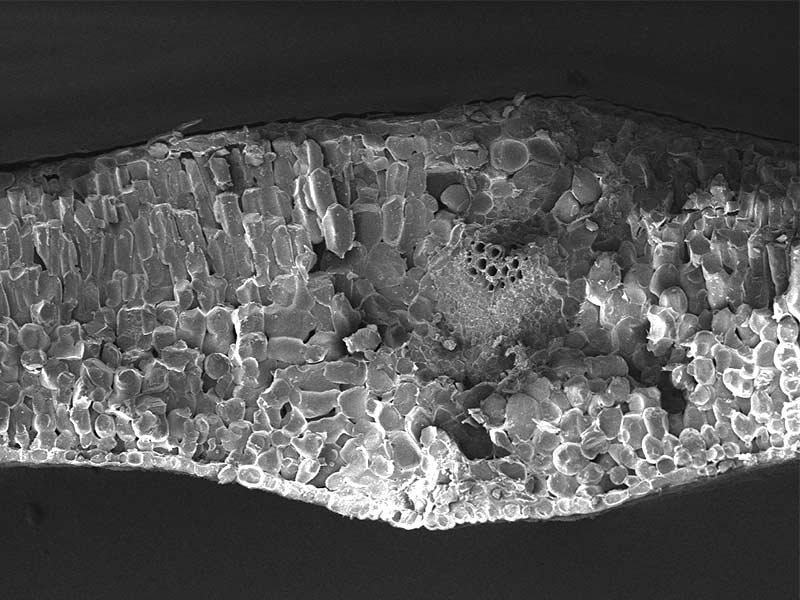
3. Cryo-SEM imaging of cryoplaned samples. Here a cut surface is viewed with the cryo-SEM method. The cut surface can be made using a cryo-ultramicrotome, where a very flat section is made through the sample preferably with a diamond knife. This is a very rewarding method to obtain ideal cross-sections through virtually any material, including soft and liquid materials such as emulsions, ice cream, biological tissues, etc. Often, cryoplaning and freeze-fracturing is combined to obtain optimal material insights. For more information, visit our Cryoplaning page.

SEM connects millimetres, microns, and nanometres, spanning six orders of magnitude

SEM at Consistence

Consistence is equipped with a Jeol 6490LA Scanning Electron Microscope with a Gatan Alto 2500 cryosystem. Combined with a cryo-ultramicrotome, these instruments offer the following methods:

  • Conventional Scanning Electron Microscopy
  • Low Vacuum Scanning Electron Microscopy
  • Cryo-Scanning Electron Microscopy
  • Cryofracturing and Cryoplaning
  • Energy Dispersive X-ray spectroscopy (EDS, EDX)

Very often we combine detailed scanning electron microscopy with Confocal Scanning Laser Microscopy and conventional light microscopy, to obtain holistic high resolution data of a limited number of ‘cornerstone’ samples for deep insights, followed by the much faster LM-imaging of higher numbers of intermediate samples to obtain the full overview. 

Such a multidisciplinary imaging approach reduces the risk of overlooking crucial features and helps to create efficient and excellent interpretation. A special service of Consistence is to translate multidisciplinary findings into schematic drawings, which in turn can be utilised to communicate a new hypothesis, patent, or processing insight.

Scanning Electron Microscopy

The SEM at Consistence can be booked for use by third parties. Consistence also provides help in optimising SEM setups in other R&D laboratories.

Drop your question here

To visit our laboratory, send a sample or learn what we can do for you.

Our aim is to provide world class microstructural insights and we are happy to receive feedback. Contact us for more information if this page is relevant for your research. Free use of the images and content of this webpage is permitted with reference to Consistence.