The 21st century is called the Age of Biology, just because there is so much to learn from nature with new technologies. We now get increasingly more detailed insights in genetics, chemistry, and structure of cells and living organisms.
Imaging expertise
Biology research has been increasingly multidisciplinar, combining many expertises to address the research questions. Microscopy and microstructure research is often an indispensable expertise to understand biological processes and interactions. Thorough microscopical research is required to understand the impact of development, stress, genotype and other influences on the functioning of organisms.
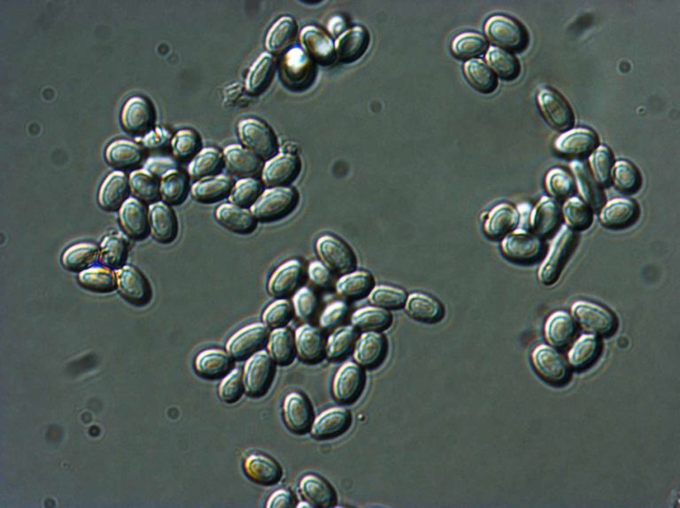
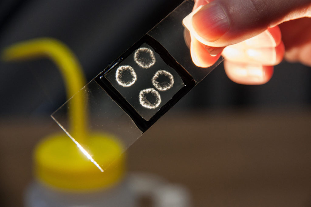
Differential Interference Contrast Microscopy (LM-DIC or Nomarski) image of fungal spores in citrus fruit. Image Width is 84µm.
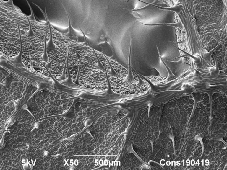
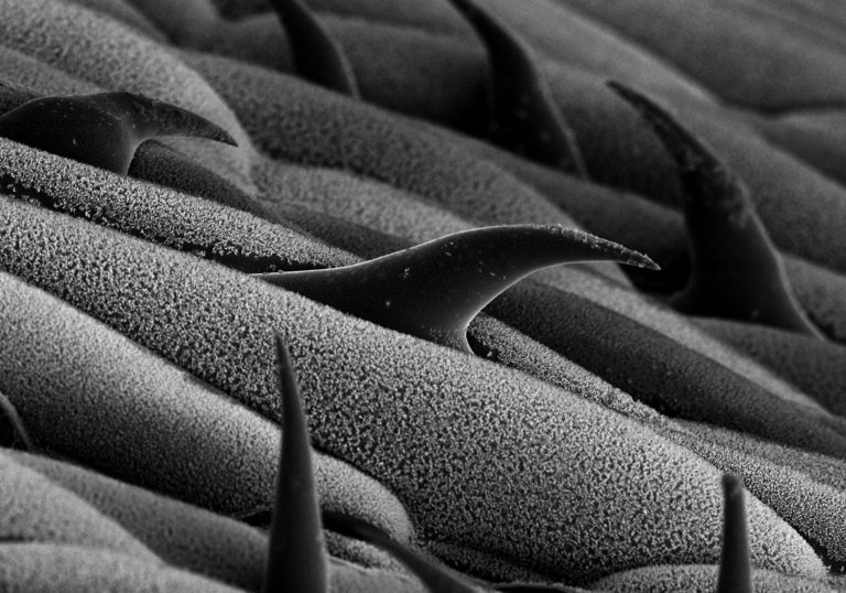
Cryo-SEM image of stinging nettle (Urtica dioica) dorsal leaf surface.
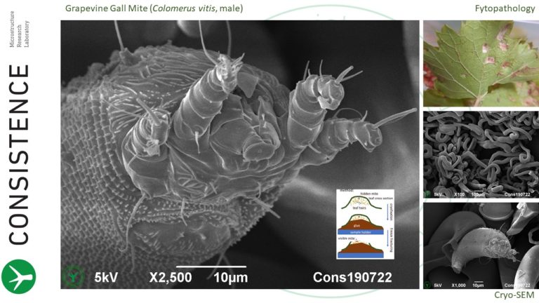
Cryo-SEM imaging of grapevine gall mite, (Colomerus vitis), hidden in leaf gall of grape vine (Vitis vinifera).
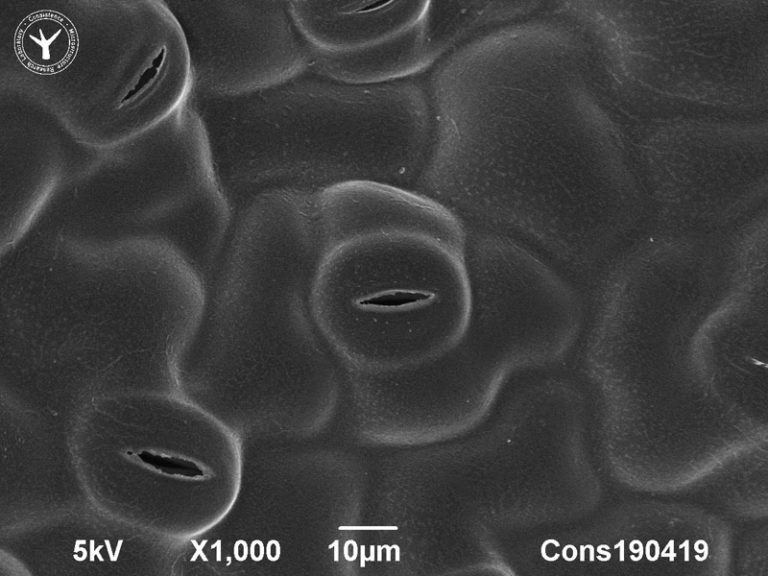
Cryo-SEM image of stomata on dorsal leaf surface of stinging nettle (Urtica dioica).
Cryo-SEM image of wax crystals on a cabbage leaf (Brassica Oleracea). The typical composition and structure of this natural wax coating creates superhydrophobicity and a special white gloss effect on cabbage leafs.
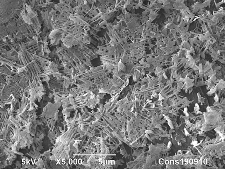
Cryoplaning SEM image of cross section through fibrous particle in cabbage puree. Image width is 120 µm.
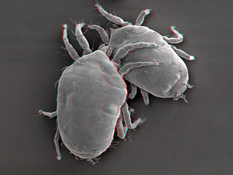
Stereo cryo-SEM image of two plant mites. Use red-green glasses to obtain 3D-view.
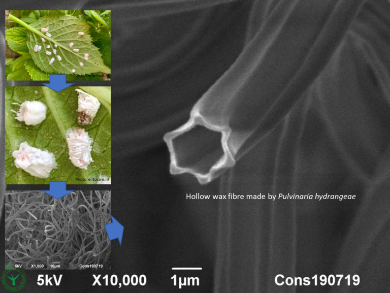
Hollow wax fibres made by hydrangea scale insects.
Cryo-SEM image of grass leaf surface. Image width is 130 µm.
Differential Interference Contrast Microscopy (LM-DIC or Nomarski) image of Chrysanthemum cut flower pith tissue revealing cell wall pits at high contrast.
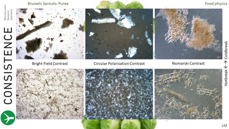
Light Microscopy analysis of hotbreak vs coldbreak brussels sprouts puree.
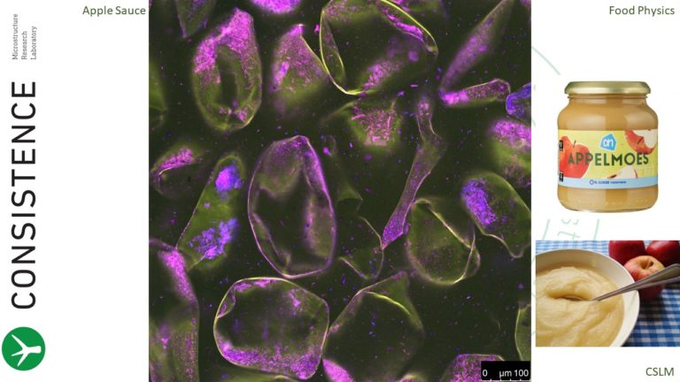
Confocal Microscopy analysis of apple sauce, with acridine orange fluorescent dye.
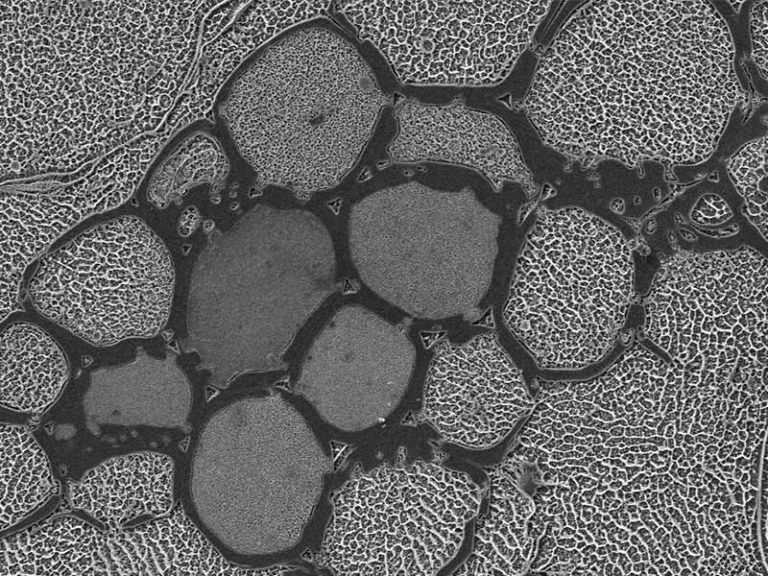
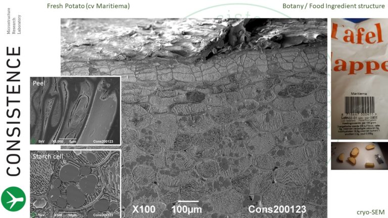
Cryoplaning SEM cross section of fresh potato.
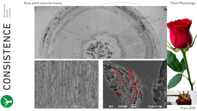
Cryoplaning SEM cross section of cut rose stem, revealing vascular occlusion by bacteria.
Living matter has stunning complexity, and astonishing order
Life Science at Consistence
Consistence has the imaging tools to study structure at multiple length scales in a single sample, from centimetres down to tens of nanometres, spanning more than six orders of magnitude. Cryofixation and subsequent cryoplaning yields large cross-sections of samples without use of any chemical fixation, revealing in situ details at nanoscale, in the larger context.
Contact us
To visit our laboratory, send a sample or learn what we can do for you.
email: info at consistence.nl
tel: +31 6 4468 1439
address: Fennaweg 53, 2991 ZA Barendrecht, The Netherlands.
Our aim is to provide world class microstructural insights and we are happy to receive feedback. Contact us for more information if this page is relevant to your research. Free use of the images and content of this webpage is permitted with reference to Consistence. Contact us for full resolution images.