template tekst – Scanning Electron Microscopy (SEM) is a method where a focussed beam of electrons is scanned over the sample surface. The recorded signal is scanned onto a screen or into a digital image. A conventional SEM requires a high vacuum sample chamber, where the electron bundle interacts with the sample. Low Vacuum and environmental SEM instruments are available as well.
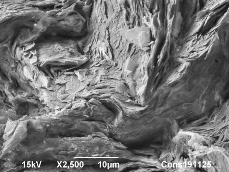
Backscattered Electron SEM image of graphite in a fractured pencil, showing higher order alignment of graphene sheets. A tilted sputtercoating with platinum was applied to obtain a shadow contrast.
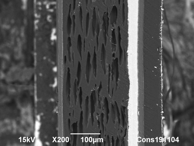
Backscattered Electron SEM image of a multilayered foil, containing an Aluminium sheet.
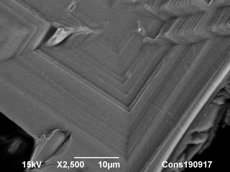
Backscattered Electron SEM image of the surface of a KCl crystal.
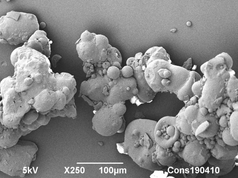
Cryo-SEM image of a commercial milk powder. Microstructure is of direct influence on powder properties.
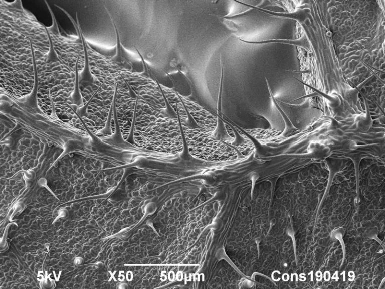
Cryo-SEM image of stinging nettle (Urtica dioica) dorsal leaf surface.
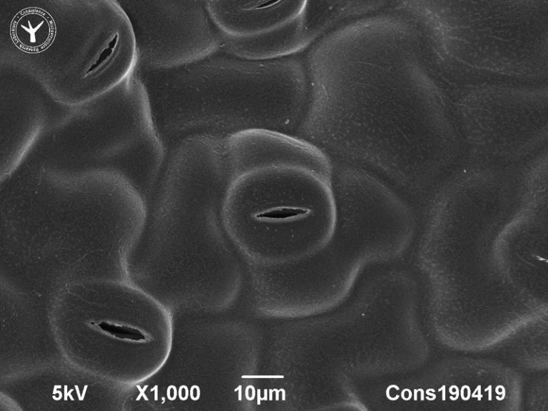
Cryo-SEM image of stomata on dorsal leaf surface of stinging nettle (Urtica dioica).
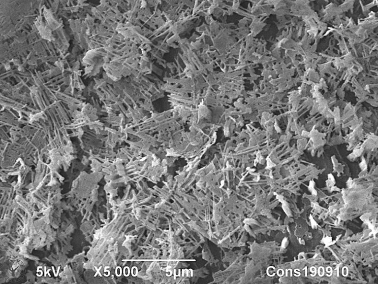
Cryo-SEM image of wax crystals on a cabbage leaf (Brassica Oleracea). The typical composition and structure of this natural wax coating creates superhydrophobicity and a special white gloss effect on cabbage leafs.
Extra titel
extra tekst Several types of electron sources have been developed to deliver optimal imaging results, either in costs or in image resolution. Different types of signals are generated by the electron bundle, of which the most used are mentioned here:
Drop your question here
To visit our laboratory, send a sample or learn what we can do for you.
Our aim is to provide world class microstructural insights and we are happy to receive feedback. Contact us for more information if this page is relevant for your research. Free use of the images and content of this webpage is permitted with reference to Consistence.