Consistence Company Video
Welcome to Consistence Microstructure Research Laboratory. The door is open to you, researchers in different fields where structure of materials, products, prototypes, biological samples, etcetera is of key interest.
Welcome to Consistence Microstructure Research Laboratory. The door is open to you, researchers in different fields where structure of materials, products, prototypes, biological samples, etcetera is of key interest.
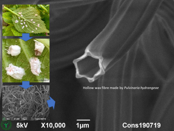
To make these hollow, hexagonal fibres would be a good challenge for nano engineers, please react! Good for them to know that the cottony hydrangea scale insects (hortensia woldopluis in dutch) already have the method in perfection. These tough, hollow fibrils protect the young scale insects from being eaten by other bugs. Magnify your own product at Consistence.
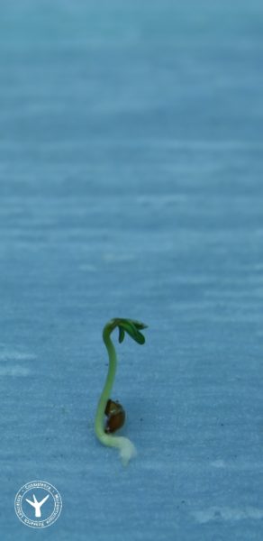
Yes, good to share that material research at Consistence is the seed to your business success, but this outburst of life, from dry seeds taken out of an aluminium sachet, makes me humbled after all...... Thanks Paul for imaging garden cress (Lepidium sativum) seed germination in our lab.
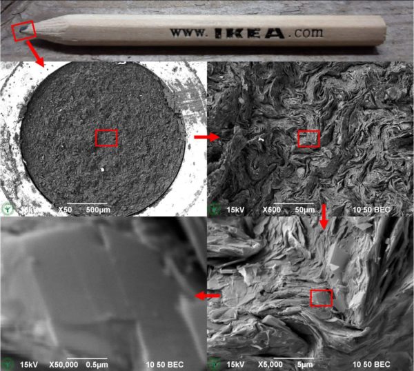
IKEA pencil magnified 50k times in four steps. High magnifications of materials and products seem disconnected from the real world. But here I would like to prove that only 4 steps are needed to magnify the graphene platelets in a pencil at a magnification of 50,000x (fifty thousand times) in just four steps, where each further zoomed image still can…
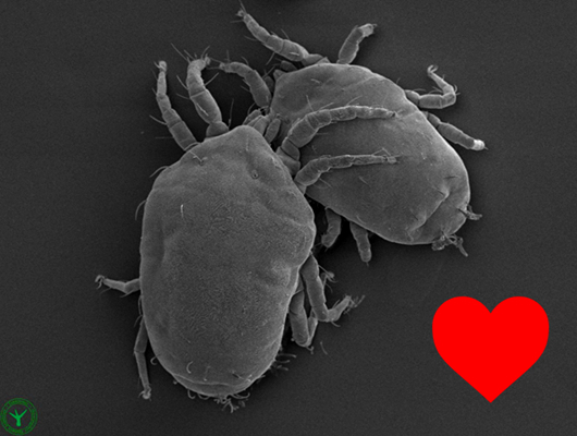
Fashion, Passion, Sports, and Defence: it's all present in the microworld ...... or just a projection of human behaviour on tiny creatures ;) The imaged mites originate from hydrangea leaves. Their weight is less than a millionth of a gram. Lowest image is a surface detail. Method: cryo-SEM. Photographed by Frank Nijsse at Consistence.
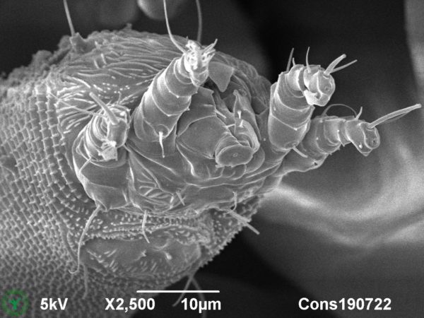
Owners of grapevines would recognise the protrusions on the imaged leaf as blisters created by the gall mite. The mites live underneath the leaves, where they have induced outgrowths of grape leave skin cells, creating a fine curly home for themselves ;). But who has ever seen the gall mite? Not so easy….. First, their size is about 0.1mm, which…

11th May Open Day at Consistence: great to see young people fascinated by science. Growing salt and sugar crystals here +imaging with polarised light.
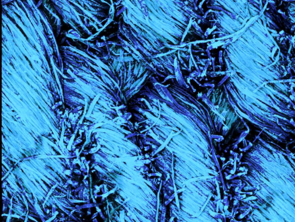
CSLM autofluorescence image of magnified worn jeans This is a confocal microscopy 3D image of denim, as an example of the amazing effect of rather low magifications of daily stuff. The image shows woven threads that are composed of individual cotton fibres. The here imaged jeans have been worn as can be concluded from the many loose cotton fibres. The…
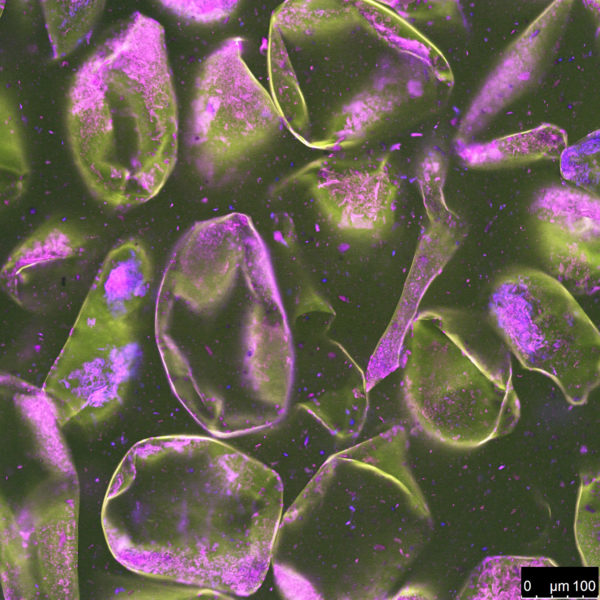
The microstructure of apple sauce. The microstructure of this beloved product is made up of loose cooked apple cells. Earlier in life, in the fresh apple, the cells were voluminous. Here they have collapsed, but they are stiff enough to hold the liquid in place and to provide the typical shear-thinning texture. The video shows a stack of confocal laser…