


Gele wratspons (Celtodoryx ciocalyptoides)
Net buiten ons normale zichtsbereik kom je niet zelden de mooiste structuren tegen. Hier zien we een gele wratspons, een exoot die in de Oosterschelde te vinden is. Mikkel Suijker fotografeerde dit dier(!) zowel onder water als in de microscoop. Het skelet van deze spons bestaat uit kunstig gevormde kiezelnaaldjes (spiculae). Qua samenstelling zijn de […]

Perception flips over
Perception flips over….You may see one star engraved in the background while the other one is on top. Actually, the two images are one and the same, just rotated 180°. This is a detail of a 10 euro cent coin. Image was made with oblique lighting and 30x magnification, creating a shadow view. Apparently, our […]

Bacterial capsules
All microscopes have their limits. Obtaining a good image at the highest resolution requires careful adjustment of microscope and sample material. But without contrast nothing would be visible. Here we pushed our tungsten cryo-SEM (Jeol 6490LA + Gatan Alto2500) to high magnifications by applying a shadowed sputtercoating of platinum, with nitrogen gas feed instead of […]

Face Mask once more
Customers at Consistence benefit from a rich combination of different imaging methodologies. And in most cases, we apply different methodologies, to reveal the various secrets of the microstructures. Here a face mask sheet, covering the filter material is shown with different light microscopic methodologies (left images) and with Scanning Electron Microscopy (right images). While one […]

Launch of SeeCHEESE project
Consistence and 3 partner companies (FrieslandCampina and 2 others) have started mapping the structure of CHEESE at all length scales, to compile the scattered knowledge of cheese microstructure. ELINE HOEDEMAKER and JANNES VAN DEN BAAR will study several cheeses with advanced imaging methods that complement each other (CLSM, SEM, cryofixed, chemfixed, etc). Combining their findings […]

Face mask cross section
In an earlier post we visualised a planar view of a face mask filter at increasing magnification, showing a heterogeneous network of fibres. It is not trivial to obtain high resolution cross sections through fabric: sub-micron (less than 1/1000 millimetre) details cannot be obtained with light microscopy or x-ray imaging, while sections of embedded material […]

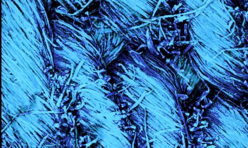
Fat crystal network
Already in 1987 Isaac Heertje published a method to visualise the fat crystal network in margarines. We now have revisited this method, making it available for today’s research of crystal networks in foods and beyond. The images show fat crystal networks in a 39% fat spread and in a 79% fat margarine, revealing continuous networks […]

Face Mask Filter
Wondered about the microstructure of a face mask filter? View here: Scanning Electron Microscope (SEM) images 33x-50kx. This disposable filter consists of melt blown polymer microfibres.

Banana browing timelapse
First a time-stack of images was acquired, resulting in the video of the ripening banana. Second, a region of interest was selected, showing the details. Third, the growing brown spots were labelled, whereby labels persisted over time, until the moment of fusion with neighbouring spots. Fourth, a ‘timesweep histogram’ was calculated, showing the brown areas growing and moving […]

Making hollow wax fibrils…….
To make these hollow, hexagonal fibres would be a good challenge for nano engineers, please react! Good for them to know that the cottony hydrangea scale insects (hortensia woldopluis in dutch) already have the method in perfection. These tough, hollow fibrils protect the young scale insects from being eaten by other bugs. Magnify your own […]

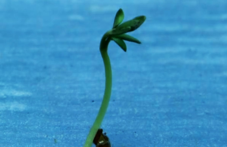
Seed germination
Yes, good to share that material research at Consistence is the seed to your business success, but this outburst of life, from dry seeds taken out of an aluminium sachet, makes me humbled after all…… Thanks Paul for imaging garden cress (Lepidium sativum) seed germination in our lab.

IKEA pencil
IKEA pencil magnified 50k times in four steps. High magnifications of materials and products seem disconnected from the real world. But here I would like to prove that only 4 steps are needed to magnify the graphene platelets in a pencil at a magnification of 50,000x (fifty thousand times) in just four steps, where each […]

Mitic Humans
Fashion, Passion, Sports, and Defence: it’s all present in the microworld …… or just a projection of human behaviour on tiny creatures 😉 The imaged mites originate from hydrangea leaves. Their weight is less than a millionth of a gram. Lowest image is a surface detail. Method: cryo-SEM. Photographed by Frank Nijsse at Consistence.

Grapevine gall mite in situ
Owners of grapevines would recognise the protrusions on the imaged leaf as blisters created by the gall mite. The mites live underneath the leaves, where they have induced outgrowths of grape leave skin cells, creating a fine curly home for themselves ;). But who has ever seen the gall mite? Not so easy….. First, their […]
Worn Jeans
This is a confocal microscopy 3D image of denim, as an example of the amazing effect of rather low magifications of daily stuff. The image shows woven threads that are composed of individual cotton fibres. The here imaged jeans have been worn as can be concluded from the many loose cotton fibres. The loose fibres […]